Glomus Tumor

Glomus Tumor

glomus tumor is a benign growth arising from the glomus body, a small thermoregulatory structure in the nail bed that regulates blood flow and temperature.

The classic presentation is severe, localized pain in a single nail with marked tenderness, often worsened by cold exposure such as cold water or air. A blue-red spot under the nail may be visible, and the lesion can cause nail thinning, ridging, or distortionBecause symptoms may be subtle and intermittent, patients often experience years of pain before diagnosis.

Diagnosis is typically confirmed with MRI or high-resolution ultrasound, which can detect the small subungual lesion. A useful clinical test is Love’s pin test, where pressing the nail with a probe produces sharp pinpoint pain directly over the tumor

Surgical excision is the treatment of choice and is usually curativeproviding immediate symptom relief. A key clinical clue is severe pain in a single nail that worsens with cold and resolves after removal of the lesion, which strongly suggests a glomus tumor. 

Pyogenic Granuloma

A pyogenic granuloma is a benign vascular growth caused by reactive capillary proliferation. Despite its name, it is neither infectious nor a true granuloma. It often develops after minor trauma, ingrown nails, pregnancy (granuloma gravidarum), or certain medications such as retinoids, indinavir, or cyclosporine.

Clinically, it appears as a rapidly growing, bright red or violaceous papule or nodule that is fragile and bleeds easily with minimal trauma. These lesions commonly occur on the nail folds and may extend under the nail plate, sometimes causing onycholysis. Because of its appearance, it may mimic conditions such as amelanotic melanoma or warts.

Treatment typically involves curettage with cautery, though cryotherapy, laser therapy, or shave excision may also be used. Treating the underlying cause, such as removing an ingrown nail spike, helps prevent recurrence. A key clinical clue is the sudden appearance of a bleeding nail fold growth after trauma or during pregnancy, which strongly suggests pyogenic granuloma.

Koenen Tumor (Periungual Fibroma)

Koenen tumor, also known as a periungual fibroma, is a benign angiofibroma that develops around the nail folds. It is strongly associated with tuberous sclerosis complex (TSC) and occurs in about 50–80% of adults with the condition.

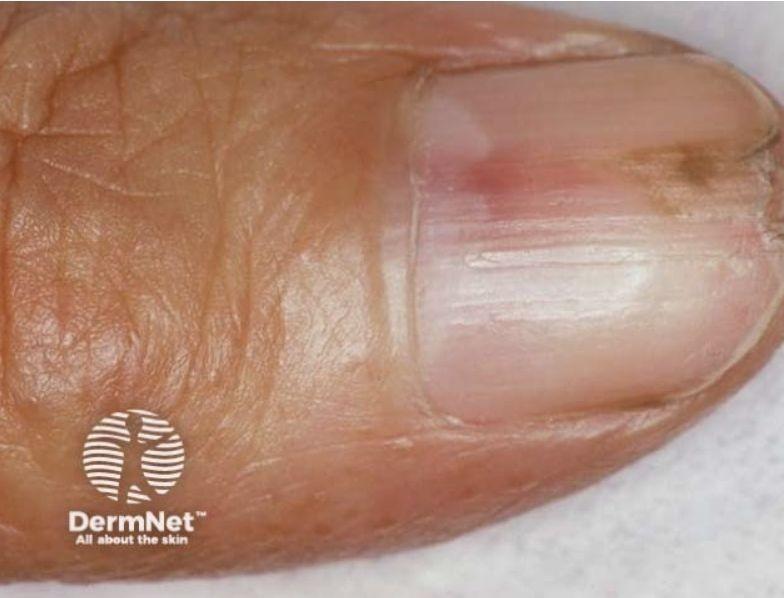
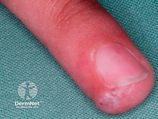
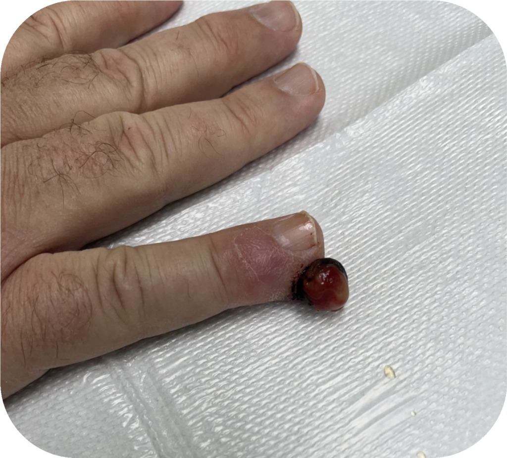
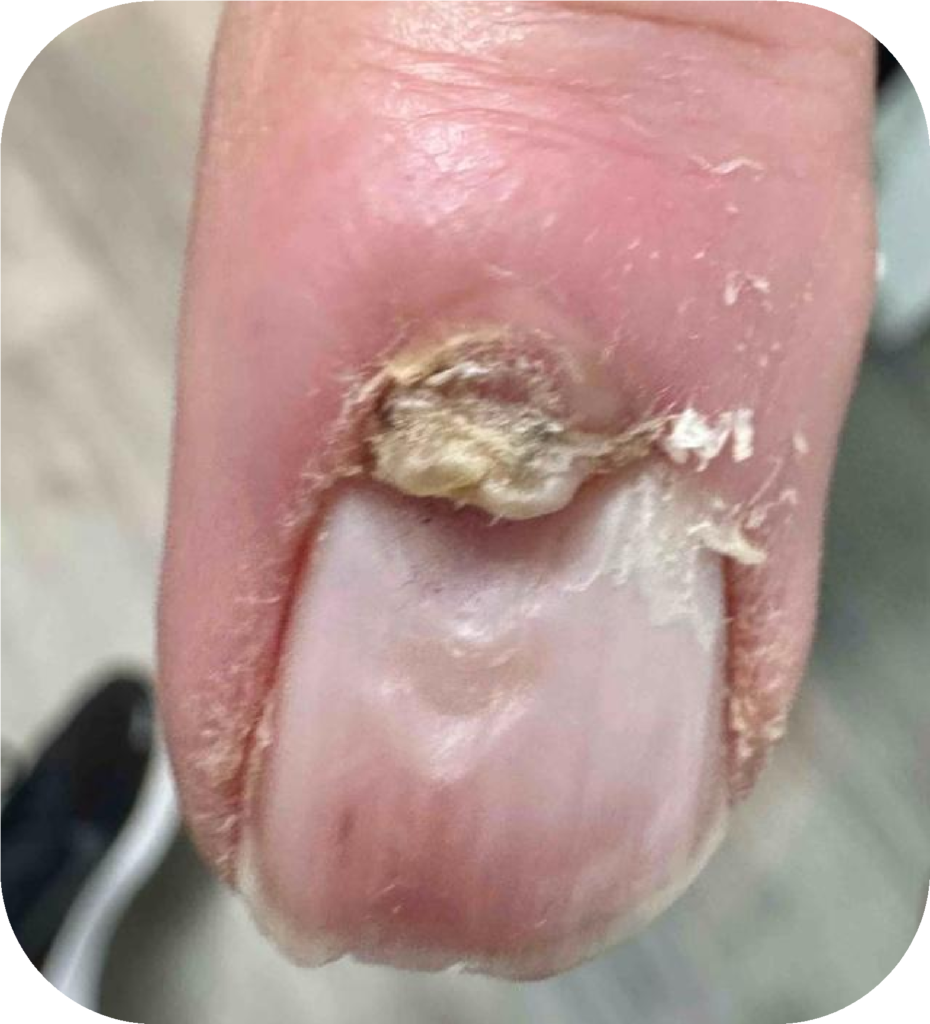
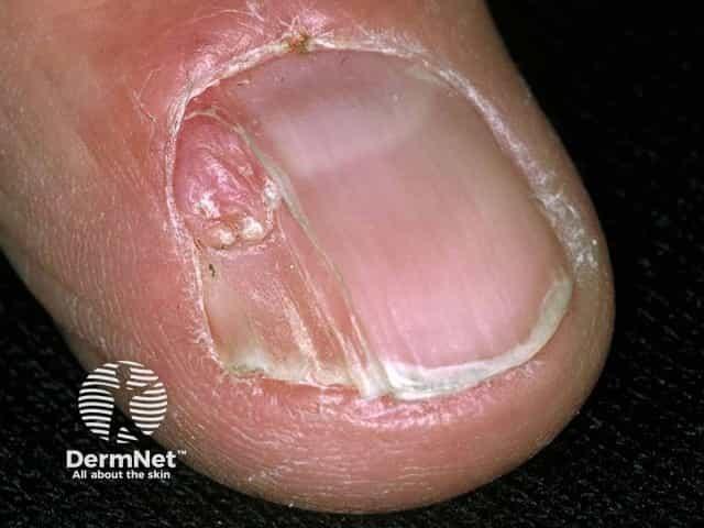
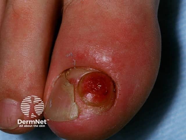
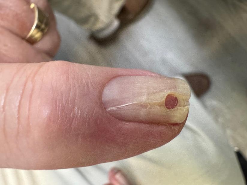
Clinically, these lesions appear as firm, smooth, skin-colored to pink nodules around the periungual or subungual nail folds. They are often multiple and may distort the nail plate, causing longitudinal grooves, ridging, or onycholysis. Koenen tumors usually develop around puberty and increase in number with age. Although often asymptomatic, they may bleed, become painful, or be easily traumatized.

Because of their strong association with tuberous sclerosis, patients should be evaluated for other features such as facial angiofibromas, hypomelanotic macules (“ash-leaf spots”), shagreen patches, seizures, and developmental delay. Diagnosis is usually clinical, though histology shows fibrous tissue with dilated vessels and spindle-shaped fibroblasts.

Treatment options include simple excision, shave removal, electrocautery, or laser ablation (CO or pulsed-dye laser)Recurrence may occur if lesions are not completely removedIn patients with tuberous sclerosis, mTOR inhibitors such as sirolimus may help reduce lesion size.

Clinical pearl: Multiple periungual fibromas in an adolescent or adult should raise strong suspicion for tuberous sclerosis complex.

Subungual Exostosis

A subungual exostosis is a benign bony growth arising from the dorsal surface of the distal phalanx beneath the nail bed. It most commonly affects the toenails, particularly the great toe.

The condition is often linked to prior trauma, repeated micro-injury, or chronic irritation or infection. The lesion consists of mature bone covered by a fibrocartilaginous cap that gradually enlarges and pushes upward against the nail.

Clinically, it presents as a firm, painful swelling beneath the nail that causes nail elevation, distortion, and sometimes onycholysis (separation of the nail from the nail bed). Pain is often worse with pressure from shoes and may make walking uncomfortable. In long-standing cases, the lesion may irritate or ulcerate the overlying nail bed.

Diagnosis is suggested by physical examination showing a hard, fixed subungual mass and confirmed with an X-ray demonstrating a bony projection from the distal phalanx.

Conservative treatment is usually ineffective. Surgical excision with removal of the bony base is the treatment of choice and is typically curative, with low recurrence when the lesion is completely removed. The prognosis is excellent since this is a benign condition that resolves after proper surgical treatment.

Myxoid Cyst

A digital myxoid (mucous) cyst is a pseudocyst filled with clear, gelatinous fluid that is usually connected to the distal interphalangeal (DIP) joint. It typically appears as a smooth, dome-shaped swelling near the proximal or lateral nail fold and is most common in middle-aged and elderly individuals, often associated with osteoarthritis.

If punctured, the cyst may release a clear, sticky, jelly-like material. Pressure from the cyst on the nail matrix often causes a longitudinal groove or depression in the nail plate.

Diagnosis is usually clinical based on appearance, and transillumination may reveal a clear cystic lesion.

Treatment options include repeated aspiration with intralesional steroid injection, cryotherapy, or sclerotherapy using agents such as phenol or polidocanol. Surgical excision or flap procedures may be required for persistent or recurrent cases.

Clinical pearl: A longitudinal nail depression with a nearby swelling should raise suspicion for a digital mucous cyst.

At Aesthetics and Medical Lasers of Colorado – Dermatology, we provide comprehensive evaluation and personalized treatment for alopecia areata and other hair disorders.

📍 Longmont Office
1350 Tulip Street, Suite 2
Longmont, CO 80501

📍 Fort Collins Office
2801 Remington Street, Suite 2
Fort Collins, CO 80525

📞 Call us today: 720-818-0533 Schedule your consultation and take the first step toward healthier, fuller-looking hair.

Popular Blog

  • What are syringomas?
  • Does sweat harm your skin
  • PH and Lipid Barrier
  • Glomus Tumor
  • Alopecia Areata
  • Female Pattern Hair Loss
  • Locations

    700 Tenacity Drive #102 Longmont, Colorado, 80504

    2801 Remington Street Suite 2, Fort. Collins, Colorado, 80525

    Contact

    720-818-0533